Structures
![](https://nadir.ch.rachip.com/wp-content/uploads/2023/07/noam-adir-seperator-green.png)
5I3A
Crystal Structure of tyrosinase from Bacillus megaterium with configuration A of hydroquinone inhibitor in the active site
![](https://nadir.ch.rachip.com/wp-content/uploads/2024/04/5I3A.gif)
8QX7
Apo-C-Terminal Domain Homolog of the Orange Carotenoid Protein from Anabaena at a resolution of 1.95 Å
![](https://nadir.ch.rachip.com/wp-content/uploads/2024/04/8QX7.gif)
8QX5
Anabaena Apo-C-Terminal Domain Homolog Of The Orange Carotenoid Protein In Native Conditions
![](https://nadir.ch.rachip.com/wp-content/uploads/2024/04/8QX5.gif)
5I38
Crystal Structure of tyrosinase from Bacillus megaterium with inhibitor kojic acid in the active site
![](https://nadir.ch.rachip.com/wp-content/uploads/2024/03/5I38.gif)
5BRT
Crystal Structure of 2-hydroxybiphenyl 3-monooxygenase from Pseudomonas azelaica with 2-hydroxybiphenyl in the active site
![](https://nadir.ch.rachip.com/wp-content/uploads/2024/03/5BRT.gif)
1R85
Crystal structure of the extracellular xylanase from Geobacillus stearothermophilus T-6 (XT6): The WT enzyme (monoclinic form) at 1.45A resolution
![](https://nadir.ch.rachip.com/wp-content/uploads/2023/08/1r85-300x300.jpeg)
1R87
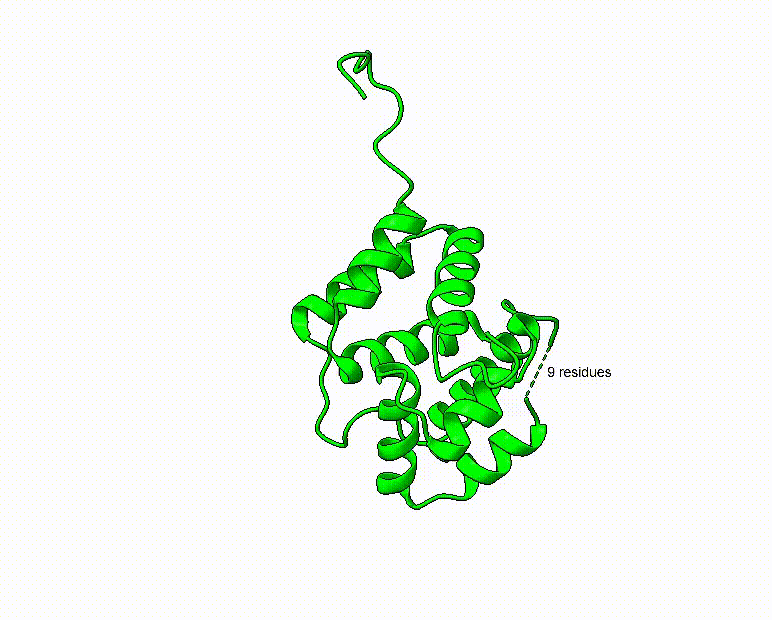
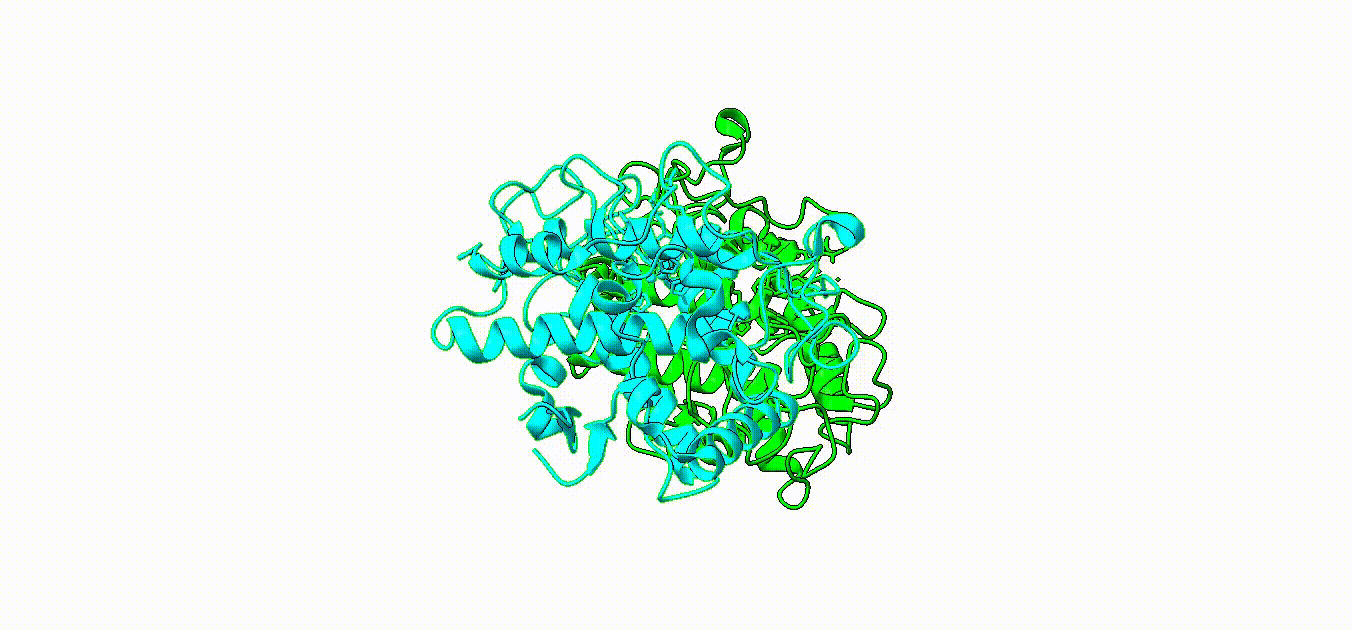
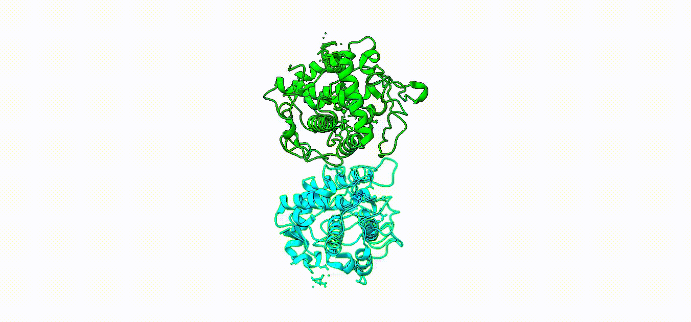
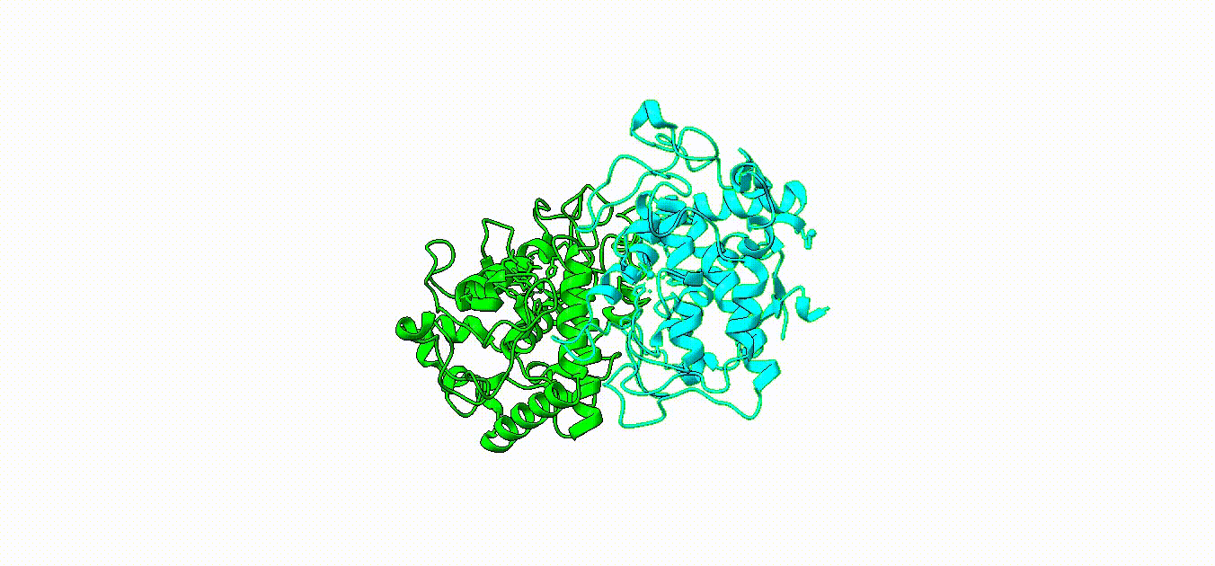
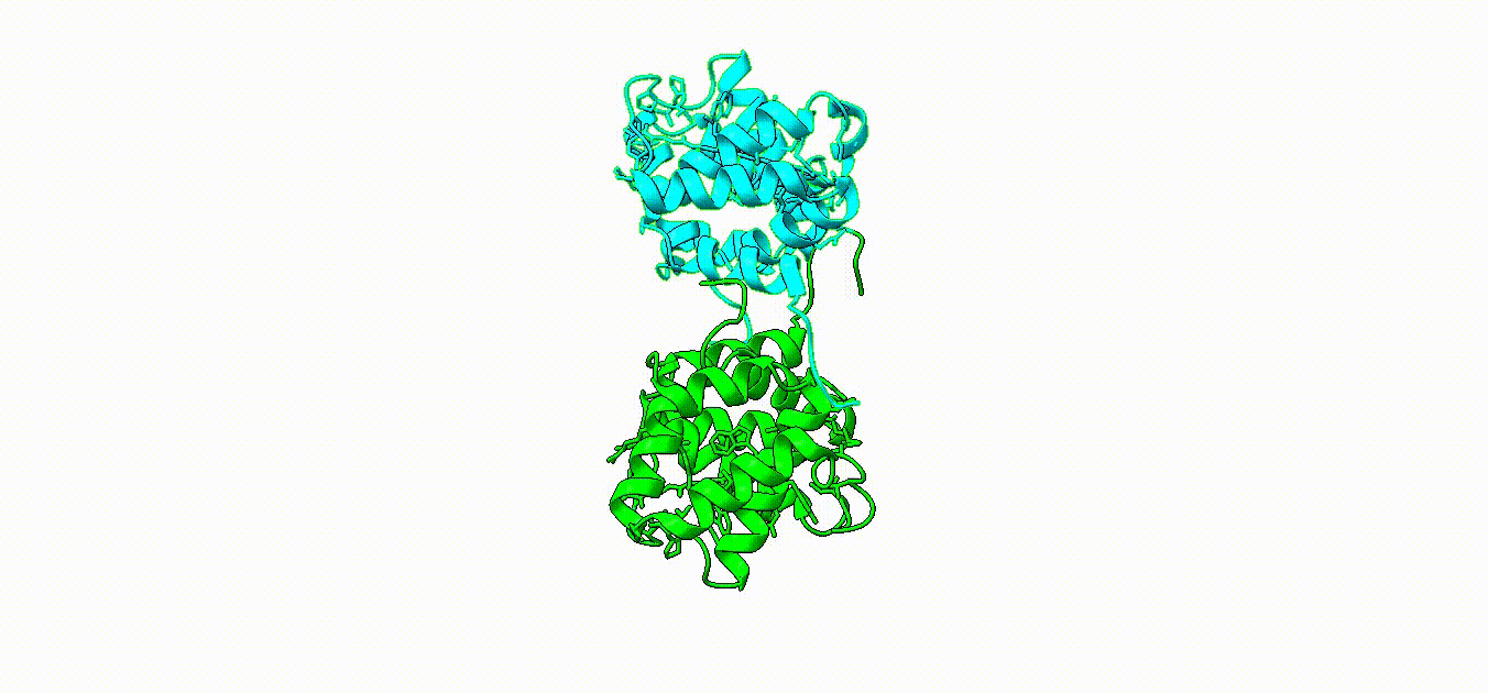
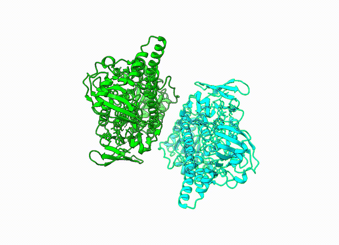
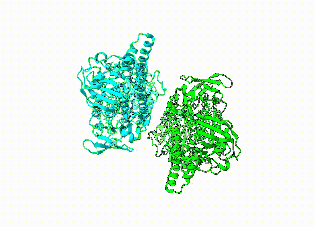
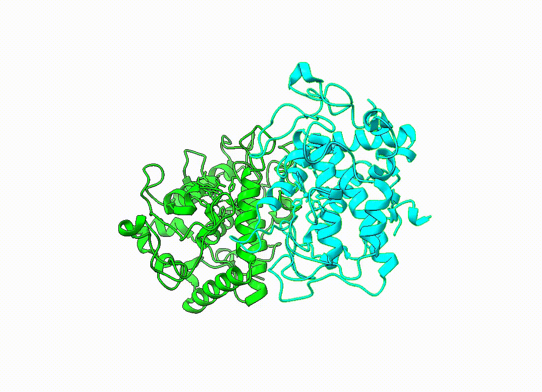
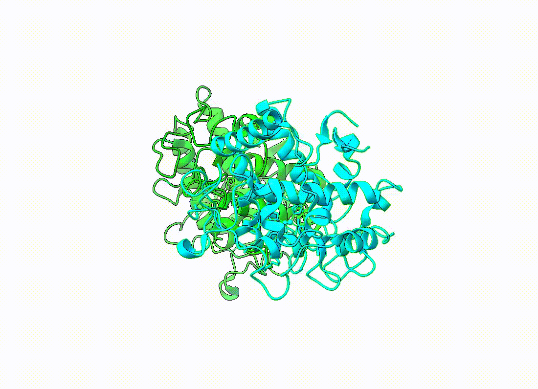
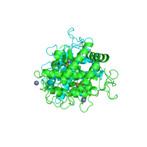
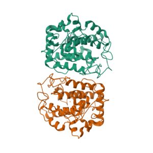
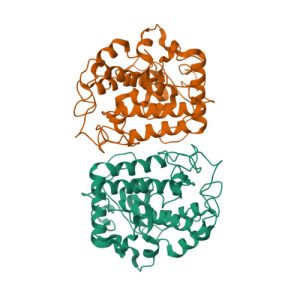
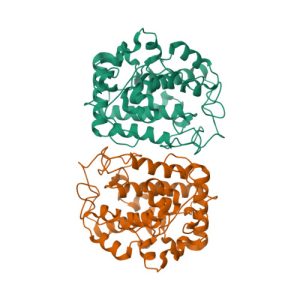
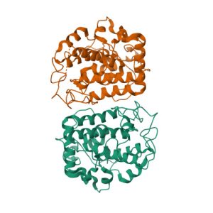
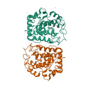
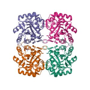
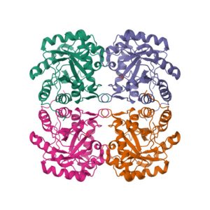
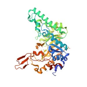
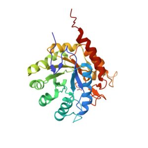
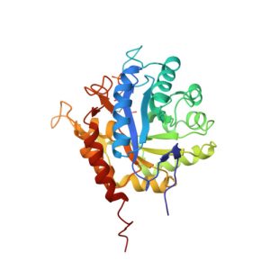
Crystal structure of the extracellular xylanase from Geobacillus stearothermophilus T-6 (XT6, monoclinic form): The complex of the WT enzyme with xylopentaose at 1.67A resolution
![](https://nadir.ch.rachip.com/wp-content/uploads/2023/08/1r87-300x300.jpeg)
1PHW
Crystal structure of KDO8P synthase in its binary complex with substrate analog 1-deoxy-A5P
![](https://nadir.ch.rachip.com/wp-content/uploads/2023/08/1phw-300x300.jpeg)
1G7U
Crystal structures of KDO8P synthase in its binary complex with substrate phosphoenol pyruvare
![](https://nadir.ch.rachip.com/wp-content/uploads/2023/08/1g7u-300x300.jpeg)
1G7V
Crystal structures of KDO8P synthase in its binary complex with the mechanism-based inhibitor
![](https://nadir.ch.rachip.com/wp-content/uploads/2023/08/1g7v-300x300.jpeg)
1I7Y
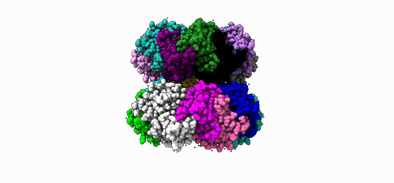
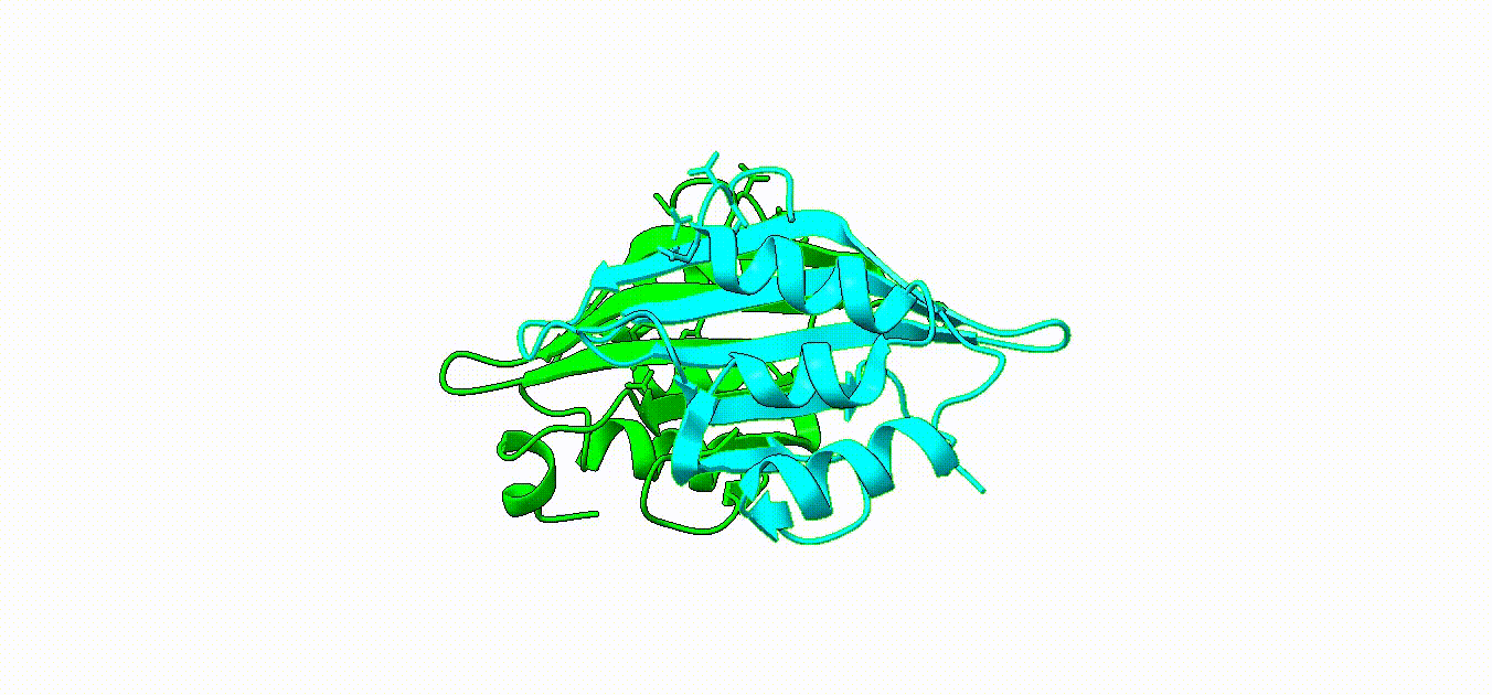
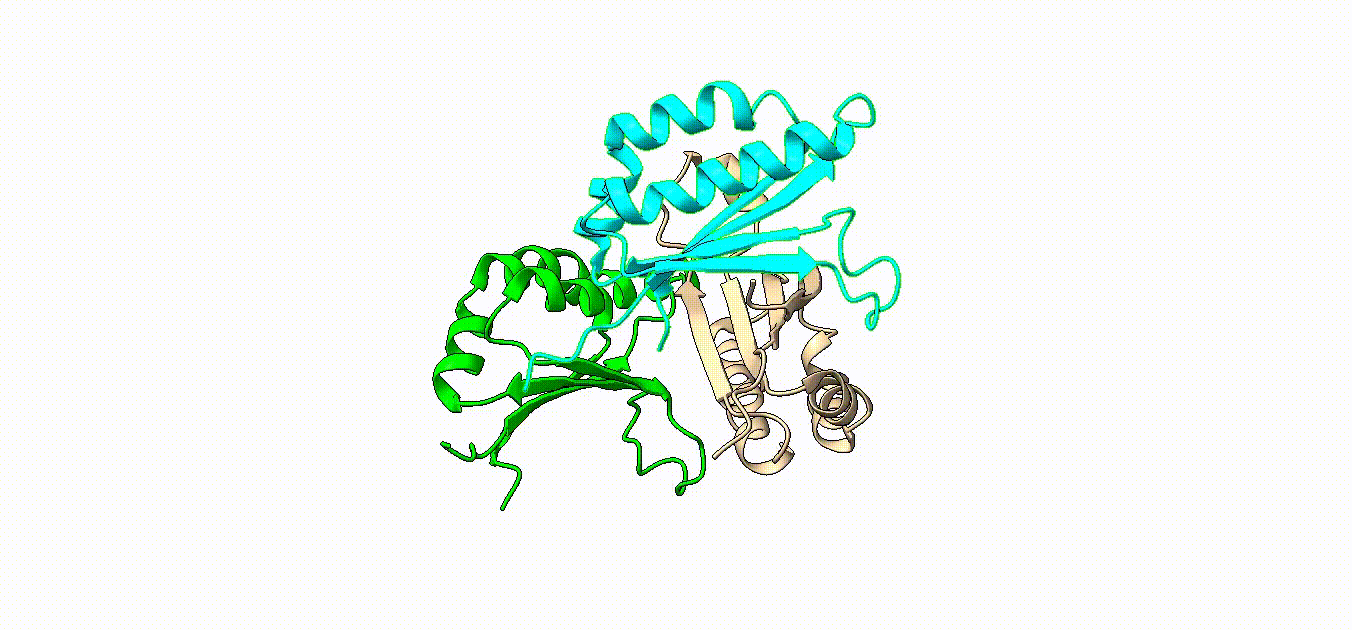
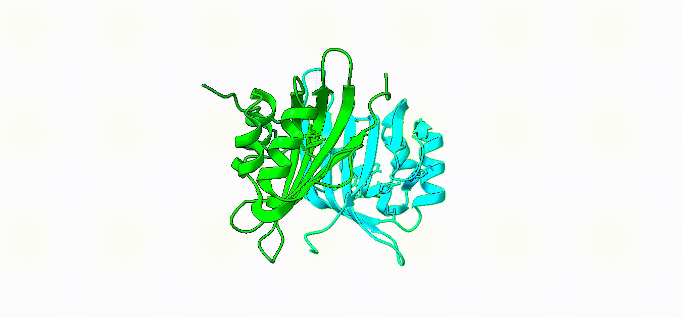
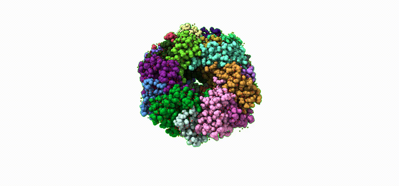
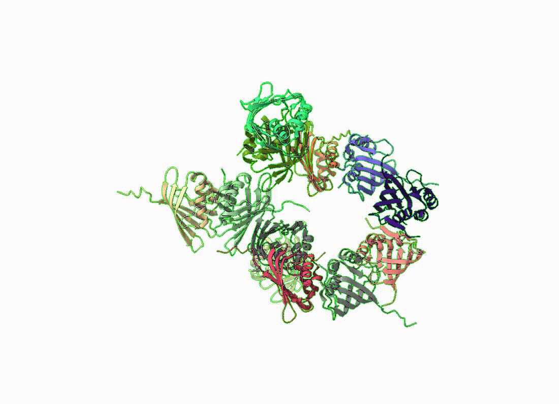
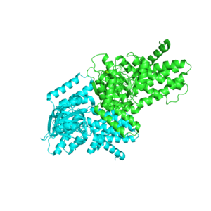
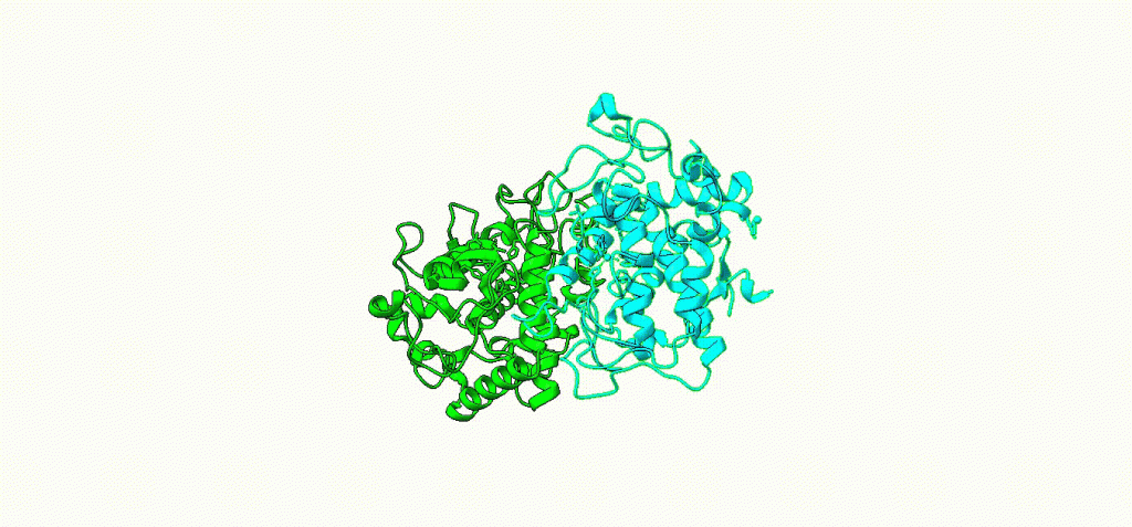
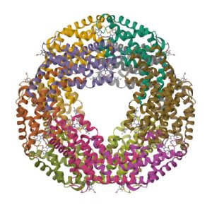
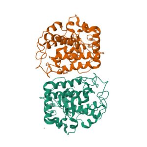
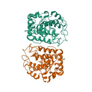
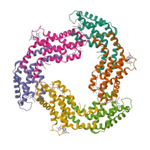
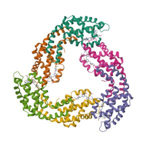
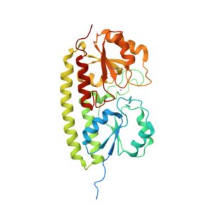
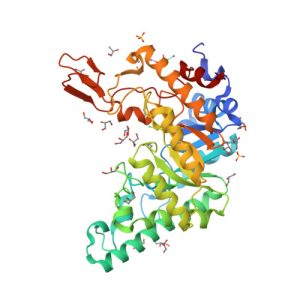
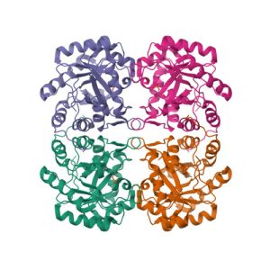
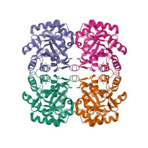
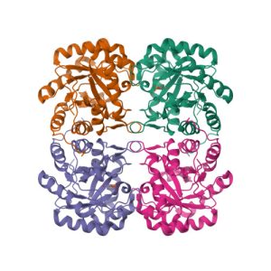
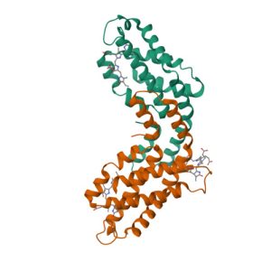
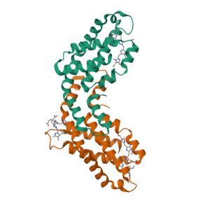
Structure of c-phycocyanin from the thermophilic cyanobacterium Synechococcus vulcanus at 2.5 A: structural implications for thermal stability in phycobilisome assembly
![](https://nadir.ch.rachip.com/wp-content/uploads/2023/08/1i7y-300x300.jpeg)